
The short answer
The original headline describes an unusual individual case report. It should not be interpreted as evidence that people without a clinical indication need brain screening.
- A case report describes one patient’s circumstances.
- Clinical history explains why imaging was performed.
- Use your own clinician’s assessment to guide examination choices.
| Look for | Ask yourself |
|---|---|
| Study type | Is this one case or a study of many people? |
| Clinical context | Why did clinicians investigate this patient? |
| Actual finding | What was demonstrated, and what remains uncertain? |
| Personal relevance | Has my clinician identified a reason for this test? |
What the original 2018 report described
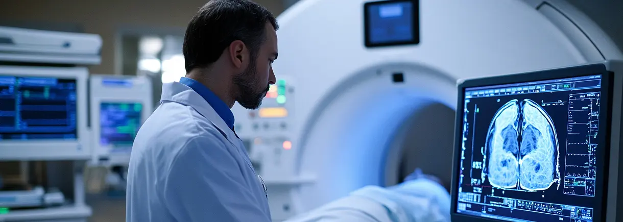
The case involved an 84-year-old man evaluated after repeated falls and new left-sided weakness. CT showed a large air-containing cavity in the right frontal region. The authors considered a bony growth near the ethmoid region and a connection through the skull base as the explanation for air entering under pressure. MRI added information, including a small adjacent area of acute infarction. Those details show why the clinical presentation and the two imaging examinations belonged together. The report concerns a particular patient and mechanism; it does not describe a routine finding in people having brain scans.
How to read a striking medical story critically
A case report can document an unusual observation without showing how often it occurs or whether screening would benefit other people. When reading a medical headline, identify the original source, the study design and the population actually described. Ask whether a conclusion is supported by one patient, a larger study or several studies that reach similar results. Different research designs answer different questions. A dramatic image may be educational, but its emotional impact is not a measure of personal risk. Bring a concern to a clinician using your own symptoms and history, rather than assuming the headline applies to you.
A case report cannot tell you how likely a finding is
A case report describes an individual clinical situation selected because it was unusual or educational. It does not provide a population estimate or show that similar symptoms usually have the same cause. A striking headline should therefore be read with the study design in mind.
If the story raises a question about your own symptoms, discuss the symptoms with a clinician rather than using the article to select a scan. The relevant history, examination and urgency determine the assessment. The lesson is the value of clinical context, not that everyone needs imaging for an uncommon condition.
Read the report with the question that prompted the scan
An imaging report may contain an observation unrelated to the reason for the examination. Your clinician can explain whether it is incidental, whether previous images help and whether any follow-up is recommended. These are separate questions from whether an observation sounds alarming.
Keep the exact wording of any report phrase you want explained. Ask which conclusion matters to your care and what action, if any, is recommended. An unusual published case should not override an interpretation made from your actual images and medical history.
Why clinical context matters
Brain MRI can help clinicians examine the brain in detail, but the choice of imaging depends on the medical question. Your own history, examination and symptoms determine the appropriate assessment. An unusual news finding should not be assumed to explain a symptom you have.
Separate the observation from the conclusion
When reading a case report, distinguish the finding shown on the scan, the authors’ explanation and the broader claims made by a headline. Those are different levels of evidence. A striking image can document an unusual event without telling readers how common it is or whether a test would help people in other circumstances. The most useful questions concern the patient’s clinical context and how the finding affected that person’s care.
Why the underlying report matters
News summaries often omit the history, examination and limitations that make a case understandable. Following the source lets you see what was actually reported and what remains uncertain. If the story prompts a personal concern, explain the concern to your clinician without assuming you have the same condition. An appropriate assessment may or may not involve imaging, and the chosen examination should answer the clinical question identified for you.
If you have a referral
Confirm the exact study ordered, the location and any preparation instructions with the imaging team. Ask your clinician how and when results will be discussed. For a medical emergency, call 911 rather than requesting a routine outpatient appointment.
Why a rare case can be informative without becoming a screening rule
Case reports can alert clinicians to unusual presentations and help document how a problem was investigated. They are not designed to estimate how often the same finding occurs in the general population or prove that a screening program improves outcomes. Keep those limits in mind when reading a dramatic headline or comparing the story with a symptom of your own.
Use the story to ask a better question
If the report raises a concern, discuss that concern with your clinician and ask what assessment is appropriate. If imaging is ordered, clarify the specific examination and the reason for it. A routine brain MRI, head CT and a vascular study are different requests. The imaging center can confirm logistics after the clinical question and order are clear.
Your Next-Step Checklist
- Distinguish an individual case from population evidence.
- Discuss symptoms rather than self-selecting a scan.
- Ask which report findings affect your care.
- Clarify any recommended follow-up.
Frequently Asked Questions
Does this case mean everyone should have a brain scan?
No. An unusual report does not establish a screening recommendation. Your own history and clinical assessment guide whether imaging is appropriate.
Where can I read the original report?
The source section links to the Brown and Vahidassr case report record. Read it in the context of the individual patient described, rather than treating the headline as general medical advice.
Did CT and MRI show exactly the same information in this case?
No. CT identified the large air-containing cavity, while subsequent MRI confirmed the finding and added evidence of a small adjacent acute infarction. That is a description of the reported case, not a rule that every person with a neurological concern needs both examinations.
Does a rare case report show that everyone should be screened for the same problem?
No. A report about one patient cannot establish how common a condition is or the benefits and harms of screening a wider population. Broader evidence is needed for those conclusions. The appropriate response to a personal concern is a clinical assessment of your own circumstances.
General educational information. Your examination and preparation depend on your clinical needs. Follow the instructions provided for your appointment.
Further reading: Brown and Vahidassr, BMJ Case Reports, 2018 · RadiologyInfo: brain MRI · medlineplus.gov: understandingmedicalresearch.html