A bone density scan is an imaging test which quantifies bone density. The lower a patient’s bone density, the greater their risk for bone fractures.
Bone density scans can be prescribed for many reasons: to predict the chance of breaking a bone, therefore preventing such an occurrence, to diagnose osteoporosis, or to see how well bone density is improving or deteriorating over time.
What is osteoporosis?
Osteoporosis, literally meaning porous bone, is a condition in which bones are too weak and bristle. Normally, there is a good balance between bone regeneration and bone destruction. If the body is losing too much bone, or not producing enough, osteoporosis can occur.
The risk factors for developing osteoporosis are: female gender, menopause, 60 years of age or older, a history of osteopenia or vitamin D deficiency, a sedentary lifestyle with poor physical exercise, low calcium diet, and certain medications such as corticosteroids.
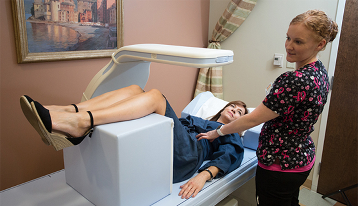
Bone density scanning:
Bone density scanning, also called densitometry, or DXA (Dual energy X-ray Absorptiometry) is a radiation-based imaging technique used to measure bone loss. It is the gold standard used to measure bone mineral density.
A DXA is a type of X-ray. As with all X-rays, a part of the body will be exposed to a small dose of radiation which will produce an image translating what’ going on in the body. Depending on the part of the body toward which the X-rays are directed, the DXA can either be central or peripheral.A central DXA is what’s commonly prescribed; it investigates bone loss in the lower spine and hip bones.
A peripheral DXA may be prescribed in some cases, or if a central DXA is not available. it will measure bone density in areas like the forearms or wrists.
T score:
The bone density test results are reported using a special international score known as the T-score.
A T score basically compares your bone density to that of a healthy 30 year old adult. When your doctor receives your bone density results, they will look at your lowest T-score and conclude from there.
According to the WHO (World Health Organization), this is what a T-score means:
A T-score of -1.0 or above is normal bone density.
A T-score between -1.0 and -2.5 means you have low bone density or osteopenia.
A T-score of -2.5 or below is a diagnosis of osteoporosis.
The lower a person’s T-score, the lower the bone density.
Osteoporosis management:
If the densitometry results indeed show the presence of osteoporosis, some lifestyle changes are necessary to manage the condition and prevent any fractures.
Primary prevention is based on weight bearing endurance exercises to increase bone density. Certain fall prevention measures need to also be taken into consideration, such as getting proper eyewear if the person is visually challenged.
For secondary prevention, in people who have already sustained fractures, certain medications (known as Bisphosphonates) can be prescribed, in addition to the lifestyle changes.