How to Deal with Back Pain

There are few conditions more debilitating than back pain. Living with chronic or even occasional back discomfort can have a significant impact on your quality of life, and might even signal more severe underlying health issues. Fortunately, there are steps you can take to minimize your discomfort, and place yourself on the road to recovery.  

What is the Source of Your Back Pain? 

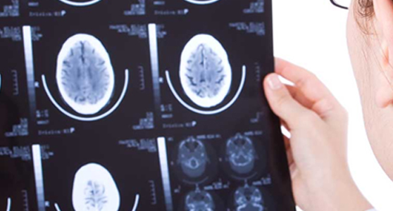
There are a myriad of causes for back pain. Some of these causes can be fairly obvious to the sufferer. At other times, the source of back pain can be more challenging to diagnose. 

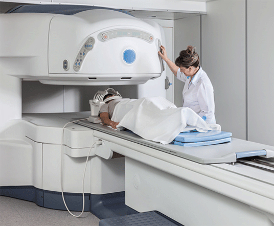
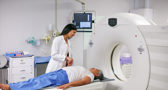
It’s a good idea to seek the counsel of a medical professional if your pain persists, increases in severity, or if you’re mystified by its root cause. Whether it’s a simple sprain, a symptom of obesity or a sedentary lifestyle, or the result of a more serious health concern, your doctor can provide you with an accurate diagnosis and course of treatment. 

Medications 

Your doctor may prescribe various brands of painkillers to help you cope with your back pain.  These medications may include prescription narcotics, muscle relaxers, or simple over-the-counter pain relievers such as acetaminophen or ibuprofen. This method of treatment should not be considered a long-term solution, however, as several of these medications may lead to dependency and work only by treating the painful effects of the condition and not necessarily the condition itself.  

Exercise 

It may seem counterintuitive to some, but in many cases the best treatment for back pain is not bed rest, but exercise. After all, many cases of back pain result from a lack of physical exertion, and healing might only be possible by strengthening the affected muscles. Gradually increasing your level of physical activity can work wonders for repairing the damaged regions in your back, improving your flexibility, and providing you with a general sense of wellbeing. 

Start slowly and learn your tolerance. Perform stretches and go on short walks until you feel you can take on more intensive activities.  

Of course, if any of the exercises you engage in cause you more discomfort, you should stop performing them immediately.  It’s important to learn the forms of exercise that best promote healing, physical and mental stamina, and enhanced flexibility given the parameters of your particular condition. Consult your physician before undergoing any exercise regimen. They may recommend less strenuous activities such as water exercises or yoga.  

Lifestyle Modifications 

Obesity is one of the most troubling epidemics facing us today.  It’s also a common culprit of back pain.  Carrying excess pounds can increase your risks for a plethora of dangerous diseases, and severely hinder your ability to lead an active and productive life.  It can also cause tremendous stress on the structure of your back. 

Diet and exercise are your greatest allies in the fight against obesity and, in many instances, back pain. As those extra pounds begin to shed, you will relieve your back of unnecessary strain and burden, and strengthen its ability to recuperate from injury. 

Your diet should arm you with the ingredients you need for muscle growth, tissue repair, and restored reserves of energy. You should avoid items that are laden with sugar, and choose foods that are rich in vitamins and proteins, such as leans meats, fresh fruits and vegetables.  Be sure to consult with your physician to determine the ideal diet regimen for you. 

Additional Therapies 

Natural supplements, such as calcium, flaxseed oil, and magnesium may also be beneficial, as might various creams or lotions that work to sooth muscles when applied. Hot and cold therapies may also be helpful in reducing inflammation, muscles spasms or tension. Electric stimulation, wherein currents of electrical energy are applied directly to the damaged nerves, has produced noticeable results for some sufferers of chronic back pain. 

With a little experimentation and under attentive medical supervision, you’ll discover which of these therapies work best for you. 

Surgery 

Considered an option of last resort, surgery may be suggested after all non-invasive means of treatment have been exhausted. Modern advancements in medical technologies have made it possible for surgeons to repair or sometimes replace damaged regions in the back in a more minimally-invasive manner than ever before.  Even so, many of these procedures still carry potential risks, and are not magical cures for every candidate.  

The most important thing to remember is that back pain does not have to be a life sentence. If you suffer from any form of back pain, you can rest assured that many solutions exist that can work for you.  

Need an MRI? It pays to shop around. Big time.

The median price difference for MRIs in hospitals versus freestanding imaging centers in the United States often varies by thousands of dollars, underscoring the need for consumers to shop around given high out of pocket health costs, data out Tuesday show.

This price spread was widest in Alaska, where the median price for hospital MRIs was $3,200 more than in imaging centers. Michigan wasn’t much better with a price difference of about $2,500.

Health care data company Amino, which released the pricing analysis, uses claims data from Medicare and up to 80% of insurers. It includes billions of health insurance claims that are 48 hours old and constantly updated.

“Most of us think there’s a big difference between $750 and $3,000 for an MRI,” says Leah Binder, CEO of the Leapfrog Group, which rates hospitals. “It’s not some minor detail on an insurance plans’s balance sheet.”

In the United States, 35 million MRIs are performed each year and $100 billion is spent on these services, about as much as is spent on cancer drugs, said David Vivero, Amino’s founder and CEO.

“Overall health care is taking over the American wallet,” said Vivero. “Our approach is to save every American money.”

Read more

by Jayne O’Donnell and Shari Rudavsky

Know Before You Go: MRI

The magnetic resonance imaging (MRI) machine is a large piece of medical equipment—intimidating for some, since your body slides inside it. You may not be sure how it works or what it does, only that your healthcare provider wants you to get an MRI study. But there’s no reason to worry. Getting an MRI is a painless process that takes less than an hour in many cases, according to radiologist Bruce Railey, MD, with Medical City Arlington in Arlington, Texas.

The MRI scan is a sophisticated imaging test healthcare providers use to help diagnose medical conditions. “It’s a cross-sectional imaging technique that uses a combination of magnetic fields and radio frequency fields to generate an image,” says Dr. Railey. One MRI test can produce dozens of images—each is called a slice—of the body part being studied.

Who needs an MRI?
Your healthcare provider may send you for an MRI for a variety of reasons. It’s mostly used to look at soft tissue like muscles and organs, as opposed to bones, according to Railey. “One of the MRI’s original applications was the imaging of the brain and spinal cord, and that is still a very common use,” he says. ”But it has also become very valuable in imaging of tendons, ligaments, joint cartilage and other soft tissue. It’s become instrumental in the imaging of the liver because it’s very sensitive in detecting if a tumor has spread from the liver to other sites.”

Before you go
Insurance companies may require a preliminary test, such as an X-ray, before they will cover an MRI. “That’s not uncommon with insurance companies, but it’s not what a physician would consider a requirement,” says Railey. “An X-ray may show an abnormality of a bone, but the MRI is far more sensitive in detecting anything with the muscles, tendons, ligaments or cartilage.”  You may need to stop eating and drinking four to six hours before the scan, depending on what body part is being scanned. For some procedures, especially tumor imaging, you may need a contrast dye administered intravenously. “The dye can increase the tissue contrast between normal and abnormal areas,” Railey says.

Read More

by PATRICK SULLIVAN

Doctors find air pocket hidden in man’s brain

(CNN) Doctors treating a patient who had complained of repeatedly losing his balance made an unexpected discovery: The 84-year-old man had a 3½-inch pocket of air in his brain.

The man had been referred to the emergency room by his primary physician in Northern Ireland.
He told his doctor about weeks of recurrent falls and three days of left-side arm and leg weakness, according to the report, published in the journal BMJ Case Reports. The patient, who is not identified in the report, did not have any visual or speech impairments and did not seem confused or have facial weakness, according to the authors.

“The thing I was most concerned about in an elderly patient with new onset limb weakness and balance disturbance was some form of stroke,” said Dr. Finlay Brown, a leading author of the report and a general practitioner in Belfast who treated the man.
The physicians performed scans of the brain to identify any signs of bleeding or brain damage caused by blocked blood vessels, according to Brown.
But what they found was much more unusual.

Small benign tumor

A computed tomography scan of the patient’s brain showed a large pocket of air — also called a pneumatocele — in the patient’s right frontal lobe that was approximately 3½ inches long.
“We knew immediately that there was something very abnormal,” Brown said. “Initially, we thought perhaps the patient hadn’t disclosed having previously had some form of operation or a congenital abnormality, but … he confirmed he hadn’t.”
The air pocket was right behind the frontal sinus and above the cribriform plate, which separates the nasal cavity from the cranial cavity.

By Mark Lieber, CNN

Read More 

MRI can help detect prostate cancer, reduce unnecessary biopsies

Written by Savannah Koplon

In a collaborative, multi-institutional study published by JAMA Oncology, researchers from the University of Alabama at Birmingham, University of Chicago, and National Cancer Institute determined that MRI-based prediction models can help reduce unnecessary biopsies in suspected prostate cancer patients and help improve personalized risk stratification.

The evidence in the study concluded that, while inclusion of MRI use as a biomarker can decrease unnecessary biopsies, it also helps maintain a high rate of diagnosis of clinically significant prostate cancers.

According to the research, the MRI-based model had a lower false-positive rate than the baseline model, and its use could result in performing 18 fewer unnecessary biopsies per 100 men.

“This research will help us optimize patient selection, determine risk-stratification more accurately and even reduce associated morbidity as it relates to prostate cancer diagnosis,” said Soroush Rais-Bahrami, M.D., assistant professor of in UAB’s Department of Urology, associate scientist in UAB’s Comprehensive Cancer Center, and co-author of the study. “Additionally, this imaging biomarker will enable physicians to provide an even more individualized approach to patient care by way of more efficient and accurate prostate cancer detection moving forward.”

Originally published on UAB News

New evidence supports mammography screening at age 40

A study to be presented at the American Roentgen Ray Society annual meeting at the end of April supports screening mammography starting at age 40.

“We showed the data of our screening population of 50 and over (as USPSTF recommends screening age 50 and over) and then showed how the data changes when we add the screening population 40-49 to the previous group (as ACR and SBI recommend screening at age 40),” Dr. Abid Irshad of the Medical University of South Carolina, told HCB News.

Irshad and his team evaluated over 41,000 screening mammography exams in the areas of callbacks and recall rates, biopsies performed, cancers detected and sensitivity and specificity. The three age groups studied were 40-49, 50-59 and 60-69.

Across all age groups, there were 5,196 callbacks, 1,164 biopsies and 326 cancers detected. Women age 40-49 account for 8,913 mammograms, and had 1,518 callbacks and 306 biopsies, while 52 cancers were detected.

In the 50-59-age group, 13,288 mammograms were performed and there were 1,659 callbacks, 371 biopsies, and 103 cancers detected. The 60-69-age group had 12,119 mammograms, 1,239 callbacks, 302 biopsies and 89 cancers detected.

“What we found was that by including the screening data of the age group 40-49 to the screening data of 50 and above, 19.3 percent additional cancers were found at the expense of an overall 1.5 percent increased callbacks and 0.1 percent increased biopsies,” said Irshad.

Read More at Dotmed.com

Originally published on Dotmed.com