The staff is Amazing especially the MRI Tech she was patient and helpful due to my anxiety.
Ciairra Washington
 Google review
Specialized imaging
Dedicated Prostate Imaging | Board-Certified Radiologists
Best Price Guarantee 4.9
Patient stories
Real patient stories from Advantis MRI & Diagnostic Imaging.
4.9 / 5from 1,000+ reviewsThe staff is Amazing especially the MRI Tech she was patient and helpful due to my anxiety.
Google reviewGreat customer service and gave me really good comfort while getting my MRI. I will recommend coming here for services.
Google reviewThey work to make sure things are comfortable and correct. Great place to get an MRI, if you have to.
Google reviewThis is my third visit to Advantis and I highly recommend them for x-ray and imaging needs.
Google reviewI've had 3 MRI's, one CT scan and one X-ray from this fine group. Very efficient and extremely friendly.
Google reviewVery clean and neat lobby and they have snacks!! Staff was wonderful and the tech was really quick and generous.
Google reviewUnderstand the examination
Prostate MRI can help clinicians evaluate the prostate and investigate suspected cancer or a known diagnosis. MRI findings are considered with PSA results, examination findings and other information. An MRI alone does not establish or rule out every prostate cancer.
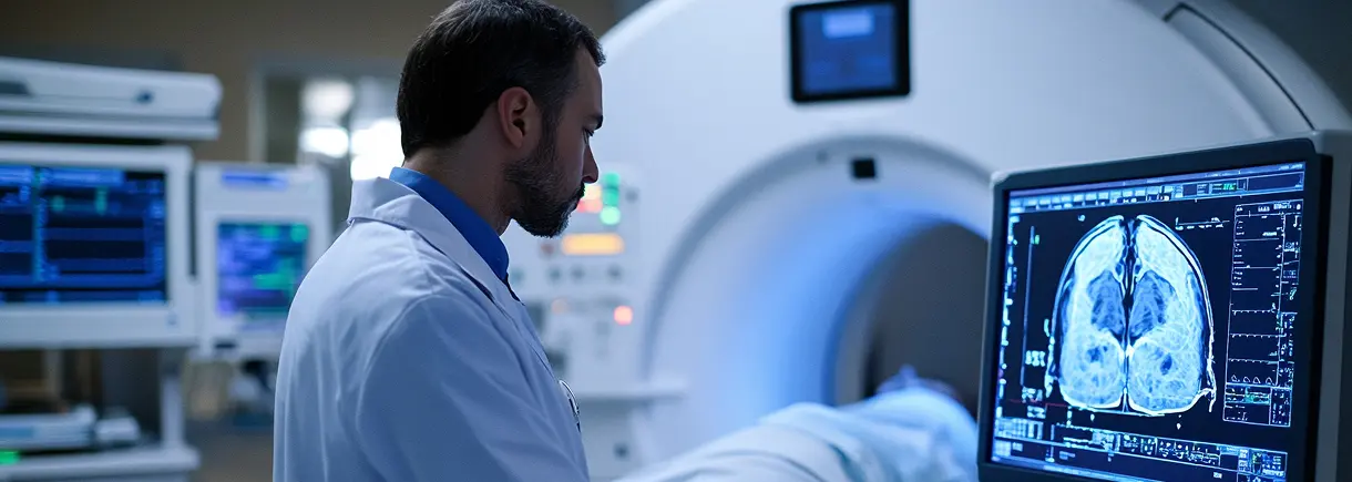
A prostate MRI uses images tailored to the gland rather than a general survey of the pelvis. Multiparametric MRI combines anatomical detail with information about water movement and, when included, how tissue behaves after contrast. Together these features help the radiologist assess suspicious areas and their extent. The examination may be requested because of a concern raised by other tests or to help assess an already known condition. Prior biopsy and treatment details matter because they can affect how the prostate looks. The protocol is chosen for the clinical question; a test labelled simply “pelvic MRI” is not automatically the same study.
A prostate MRI report may assign a PI-RADS category from one to five to an area being assessed for clinically significant cancer. Lower categories indicate lower imaging suspicion; a category of three is indeterminate, and higher categories indicate greater suspicion. This is different from grading cancer in tissue obtained at biopsy. The report may also describe the location and size of a finding, the appearance outside the gland and any limitations of the examination. Discuss the category alongside the reason the scan was ordered. A single number should not be separated from the written impression and the clinician’s assessment of the broader picture.
MRI can identify an area that deserves closer assessment and can help guide targeted biopsy. In an MRI–ultrasound fusion approach, images from the MRI are combined with ultrasound guidance to direct tissue sampling. That is a separate procedure from the MRI examination itself. Research supported by the National Cancer Institute has examined using targeted sampling together with systematic sampling, because the techniques can provide complementary information. For patients, the practical question is how the scan result changes the next decision: whether additional assessment is needed, whether a target has been identified and how the clinician will consider the possibility of disease outside that target.
PSA is produced by both normal and malignant prostate cells. An increased blood level can occur with cancer, but also with noncancerous enlargement or inflammation. The National Cancer Institute notes that there is no single PSA value that proves the presence or absence of prostate cancer. This helps explain why clinicians may review a trend, repeat testing or use other information before deciding on MRI or biopsy. Medication use and recent events that can affect the result may also matter. Keep a dated record of previous results and the circumstances in which they were obtained. The purpose of an MRI after a concerning PSA result is to add information to that evaluation, not to reinterpret the blood test as a diagnosis by itself.
If biopsy is recommended, tissue is collected for examination by a pathologist. Sampling may be performed through different approaches, and image guidance helps direct the procedure. The biopsy process has its own consent, preparation and aftercare, which differ from a diagnostic MRI appointment. The radiology report and the pathology report also have different purposes: one describes imaging observations and the other describes the tissue that was sampled. A useful consultation explains why sampling is recommended, which areas will be assessed and how results will be combined. It should also explain the limitations of sampling and the next step if clinical concern persists. This is general background on the pathway, not a statement that a particular biopsy technique is available at Advantis.
A prostate MRI may be interpreted differently when there has been a recent biopsy, previous treatment or known prostate disease. Give the treating team the dates and reports from those events, rather than only a summary that a prior test was normal. Explain whether the purpose is an initial assessment or a later evaluation of an established problem. The radiologist can then relate image appearances to that history and describe any limitations that affect the clinical question.
| Focus | What the study can add | What to keep in mind |
|---|---|---|
| Prostate tissue | Dedicated MRI can examine areas within the prostate in detail. | Its role depends on the wider urological assessment. |
| Clinical information | PSA history, examination findings and earlier procedures can inform interpretation. | Imaging should not be interpreted apart from that context. |
| Next steps | Results may help a clinician plan further investigation. | MRI alone does not establish every diagnosis or eliminate every need for biopsy. |
A doctor’s order is required before your scan. Let the team know about previous biopsies, prostate procedures, implants and prior imaging. Ask which records the radiologist needs and whether your examination requires contrast or additional preparation.
This dedicated examination is arranged at the Dickinson center using High-field MRI. MRI appointments are scheduled on Thursdays. Confirm the current availability, preparation and exact study when you call; this examination is not booked at Richmond Avenue or on the Walters Road open scanner.
The team explains positioning and communication for the scan. Follow instructions about staying still and any brief breathing steps. Ask before the scan begins how to raise a concern, and tell the technologist if you need help.
Your radiologist’s report goes to the ordering clinician, who will explain the findings and next steps. Contact our records team for copies of your images or report.
Request images and reportsRequest an estimate for the examination on your referral, including contrast and radiologist interpretation. Confirm your plan’s network status and authorization requirements.
Costs, insurance & financingAvailable locations: Dickinson. Confirm your exact protocol when booking.
View locations & hoursCommon questions
Sometimes MRI helps guide a biopsy decision or target a suspicious area. Your urologist decides whether biopsy is still needed based on your full assessment.
Call 877-345-4244 with your order or request a callback. The team confirms the requested protocol, appropriate center and appointment options. A callback request does not itself confirm a booking.
Ask for a current quote for the exact study, including any contrast and the radiologist’s interpretation. Confirm separate charges and payment terms. If using insurance, check your plan’s network, benefits and authorization requirements.
Read each requested study to scheduling. Ask whether separate appointments or estimates are needed and whether any preparation instructions conflict. The office can obtain clarification from the referring clinician when necessary.
Ask which earlier images and reports are useful and how to transfer them. An existing scan may help comparison, but the clinical team determines whether it answers the current question or meets any specialized analysis requirements.
Arrange the report discussion with the clinician who ordered the examination or manages your care. Ask how the report will reach that office and when follow-up is appropriate. Records staff can help arrange copies for another provider.
No. PI-RADS 3 means the imaging assessment is indeterminate for clinically significant cancer. It is neither a confirmed diagnosis nor an instruction to ignore the finding. Your clinician considers the report with the reason for imaging and your other clinical information before recommending follow-up or tissue sampling. Ask what additional information would change that recommendation and what the planned follow-up involves.
No. MRI records images, while a biopsy removes tissue for examination. A targeted biopsy may use previously acquired MRI images to identify areas for sampling, including through MRI–ultrasound fusion. Completing an MRI does not mean a biopsy has been performed or that tissue has been assessed. If tissue sampling is recommended, the clinician should explain the separate procedure and how the MRI will inform it.
Yes. Noncancerous conditions, including inflammation and enlargement, can raise PSA. The result needs interpretation with the clinical history rather than a single universal cutoff. Your clinician may recommend repeat testing or additional assessment depending on the circumstances. Do not assume either that an elevation proves cancer or that a possible benign explanation makes follow-up unnecessary.
A pathologist examines the tissue samples. The treating clinician considers those findings with imaging and other clinical information when explaining the diagnosis and options. The radiologist’s MRI assessment and the biopsy result are different parts of that process. Keep both reports and ask how the sampled areas relate to the MRI findings.
Yes. Advantis requires a doctor’s order for every scan, including self-pay appointments. You can request a callback before you have the order; it must be available before the examination.
Have your referral ready. Our team will confirm your scan, location and available appointment times.
