The staff is Amazing especially the MRI Tech she was patient and helpful due to my anxiety.
Ciairra Washington
 Google review
Specialized imaging
Brain White Matter Imaging | Diffusion MRI Analysis
Best Price Guarantee 4.9
4.9Patient stories
Real patient stories from Advantis MRI & Diagnostic Imaging.
4.9 / 5from 1,000+ reviewsThe staff is Amazing especially the MRI Tech she was patient and helpful due to my anxiety.
Google reviewGreat customer service and gave me really good comfort while getting my MRI. I will recommend coming here for services.
Google reviewThey work to make sure things are comfortable and correct. Great place to get an MRI, if you have to.
Google reviewThis is my third visit to Advantis and I highly recommend them for x-ray and imaging needs.
Google reviewI've had 3 MRI's, one CT scan and one X-ray from this fine group. Very efficient and extremely friendly.
Google reviewVery clean and neat lobby and they have snacks!! Staff was wonderful and the tech was really quick and generous.
Google reviewUnderstand the examination
Diffusion tensor imaging, or DTI, is an MRI technique that can depict the paths of nerve fibers in brain white matter. The usefulness and interpretation of the images depend on the clinical situation and the protocol used.
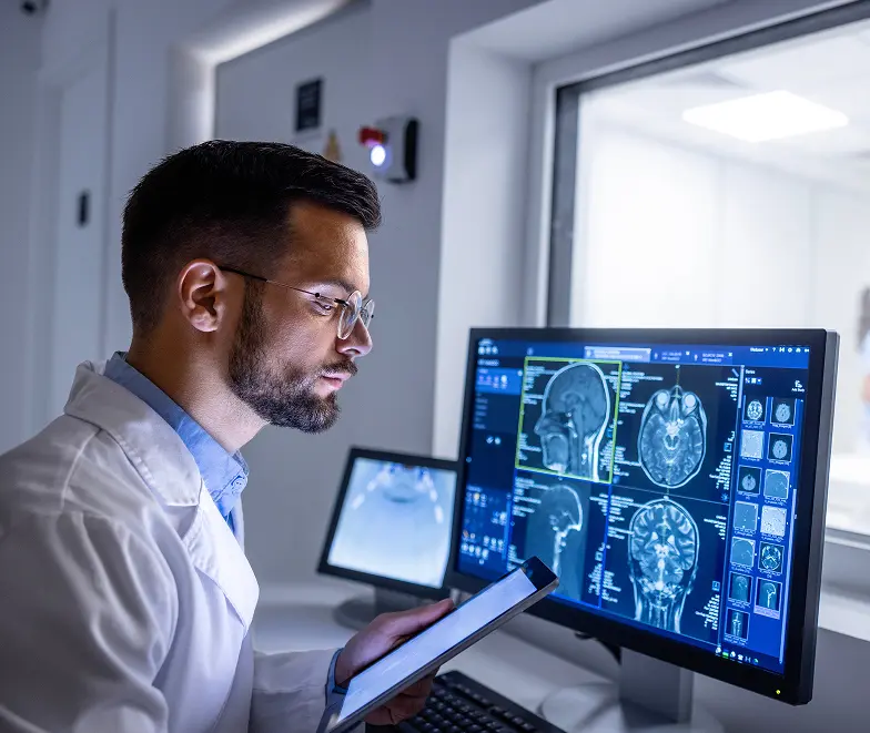
Diffusion tensor imaging examines patterns in the movement of water molecules and can be used to depict white-matter pathways. It is a particular MRI technique rather than a separate photograph of every nerve fiber. For a patient, the useful starting point is the question the specialist hopes the extra information will answer. Ask whether the request involves pathway visualization, quantitative measurements or another defined purpose, and how it will be considered alongside the conventional MRI. The images can look striking, but a colorful display alone does not explain symptoms or establish a diagnosis. The written interpretation and clinical context remain essential to understanding the result.
Tractography uses diffusion information and a computational method to estimate pathways. Research evaluating tractography has demonstrated that reconstructed connections can include false positives and can fail to reproduce the complete underlying anatomy. The result should therefore be interpreted as a model-based representation, not an exact wiring diagram. This matters when a patient sees a difference between two images or a dramatic change in color. Ask what was actually measured, what the reconstruction method can establish and which uncertainties the interpreting clinician considers relevant. A useful explanation distinguishes the observed imaging data from the conclusions that can reasonably be drawn for the individual patient.
The National Institute of Neurological Disorders and Stroke describes advances in techniques that examine subtle brain changes, including DTI, while emphasizing that current imaging methods remain inadequate for diagnosing mild concussive injury on their own. Clinical assessment includes the history of the event, symptoms and examination rather than a single scan result. A normal image does not necessarily resolve persistent symptoms, and an imaging difference does not independently establish why those symptoms occurred. Discuss how any proposed advanced imaging would change your assessment or management. The purpose should be clinically understandable rather than a promise that one technique will prove or disprove every brain injury.
Functional MRI examines small blood-flow changes associated with brain activity. It may be used to help identify regions involved in functions such as language or movement and can contribute to treatment planning in selected circumstances. DTI instead concerns diffusion information and white-matter pathways. Both can produce detailed-looking brain maps, which makes the terminology easy to confuse, but they do not measure the same thing. If a specialist requests “brain mapping,” ask which technique is meant, what the person needs to do during the examination and which decision the information will support. A task-based functional study may have requirements that do not apply to DTI. Neither label should be substituted for the other when reviewing an order.
A brain MRI can assess structures and signal changes associated with a wide range of clinical concerns, including tumors, stroke-related changes, inflammation and other neurological problems. The sequences are selected for the reason the study was requested. Advanced analysis does not make those underlying images irrelevant. In an examination that includes DTI, a clinician may need to consider conventional findings together with the additional diffusion information. Ask whether the report addresses the original symptom or referral question and whether the advanced component adds a distinct observation. The aim is an integrated interpretation, rather than two disconnected reports in which one contains ordinary images and the other contains colorful maps with no explanation of their relationship.
A request for DTI can be discussed in terms of the output needed: a pathway depiction, a quantitative observation or information for a particular specialist question. These are more useful descriptions than saying only that the scan should be “advanced.” Ask what the result can change in the assessment and which uncertainties will remain afterward. The answer should connect the technique to the clinical purpose, without promising that the appearance of a map explains every neurological symptom.
| Focus | What the study can add | What to keep in mind |
|---|---|---|
| Diffusion patterns | DTI uses MRI measurements of water diffusion. | It is a specialized technique rather than an ordinary scan label. |
| White-matter pathways | Processing can help depict pathways within the brain. | The output depends on acquisition, processing and clinical context. |
| Interpretation | A specialist relates the findings to the clinical question and other imaging. | A pathway image or measurement is not a diagnosis by itself. |
A doctor’s order is required before your scan. Ask your referring specialist what question the study is intended to answer and whether additional MRI sequences are needed. Confirm the available protocol, reporting arrangements and location with Advantis.
Contact scheduling for Richmond Avenue or Dickinson for the protocol on your order. This examination is not booked on the Walters Road open scanner. Dickinson MRI appointments are scheduled on Thursdays; confirm the next available date directly.
The team explains positioning and communication for the scan. Follow instructions about staying still and any brief breathing steps. Ask before the scan begins how to raise a concern, and tell the technologist if you need help.
Your radiologist’s report goes to the ordering clinician, who will explain the findings and next steps. Contact our records team for copies of your images or report.
Request images and reportsRequest an estimate for the examination on your referral, including contrast and radiologist interpretation. Confirm your plan’s network status and authorization requirements.
Costs, insurance & financingAvailable locations: Richmond Avenue, Dickinson. Confirm your exact protocol when booking.
View locations & hoursCommon questions
Do not assume it is. DTI is a specific technique; the order and protocol need to be discussed with the imaging team.
Call 877-345-4244 with your order or request a callback. The team confirms the requested protocol, appropriate center and appointment options. A callback request does not itself confirm a booking.
Ask for a current quote for the exact study, including any contrast and the radiologist’s interpretation. Confirm separate charges and payment terms. If using insurance, check your plan’s network, benefits and authorization requirements.
Read each requested study to scheduling. Ask whether separate appointments or estimates are needed and whether any preparation instructions conflict. The office can obtain clarification from the referring clinician when necessary.
Ask which earlier images and reports are useful and how to transfer them. An existing scan may help comparison, but the clinical team determines whether it answers the current question or meets any specialized analysis requirements.
Arrange the report discussion with the clinician who ordered the examination or manages your care. Ask how the report will reach that office and when follow-up is appropriate. Records staff can help arrange copies for another provider.
No. Tractography displays estimated pathways reconstructed from diffusion information. Its appearance depends on the data and the reconstruction approach, and it can contain misleading or incomplete connections. Ask the radiologist what the display represents and which findings are reliable enough to affect the clinical question. A picture should be considered with the written interpretation, rather than judged by color or visual complexity alone.
It should not be described that way. NINDS notes the limitations of current imaging for identifying the changes involved in mild concussive injury. Clinical history and assessment remain important even when imaging is obtained. Ask the specialist what additional information the proposed study might provide and what it cannot establish. Persistent symptoms deserve appropriate clinical follow-up regardless of whether a scan appears normal.
Task instructions are commonly associated with some functional MRI examinations, which are different from DTI. Do not assume that every type of brain mapping involves the same activities. Ask which technique has been requested and what participation it requires. If following tasks would be difficult, explain that when the examination is being planned.
It should not be assumed to do so. Different sequences provide different kinds of information, and the radiologist selects an examination for the clinical question. DTI may be one component of a broader assessment. Ask how the additional technique fits with conventional imaging and how the findings will be combined in the interpretation.
Yes. Advantis requires a doctor’s order for every scan, including self-pay appointments. You can request a callback before you have the order; it must be available before the examination.
Have your referral ready. Our team will confirm your scan, location and available appointment times.
The patient guide
Vascular & specialized imaging
DTI examines water-diffusion patterns in brain tissue. Learn how it differs from routine MRI and why interpretation needs clinical context.
MRI guidance
Understand the purpose of a head MRI, the questions to ask before booking and how to prepare for the results conversation.
