This is my third visit to Advantis and I highly recommend them for x-ray and imaging needs.
Riah Kindle
 Google review
Everyday imaging
Real-Time Diagnostic Imaging | No Ionizing Radiation
Best Price Guarantee 4.9
Patient stories
Real patient stories from Advantis MRI & Diagnostic Imaging.
4.9 / 5from 1,000+ reviewsThis is my third visit to Advantis and I highly recommend them for x-ray and imaging needs.
Google reviewThe healthcare provider performing my ultrasound was also very professional and mindful of my comfort and privacy. Thank you.
Google reviewThe staff is so amazingly nice! The receptionist is very nice and welcoming, the ultrasound technician is the best!
Google reviewThe ladies @ the front desk were wonderful and the gentleman performing the ultrasound was extremely personable. Extremely nice, and made me feel real comfortable.
Google reviewVery professional, courteous, and friendly. The ultrasound technician was really cool.
Google reviewThe staff is Amazing especially the MRI Tech she was patient and helpful due to my anxiety.
Google reviewUnderstand the examination
Ultrasound uses sound waves to produce images without ionizing radiation. Depending on the ordered study, it can assess organs, soft tissue and blood flow. Diagnostic ultrasound is a medical examination with a clinical question to answer.
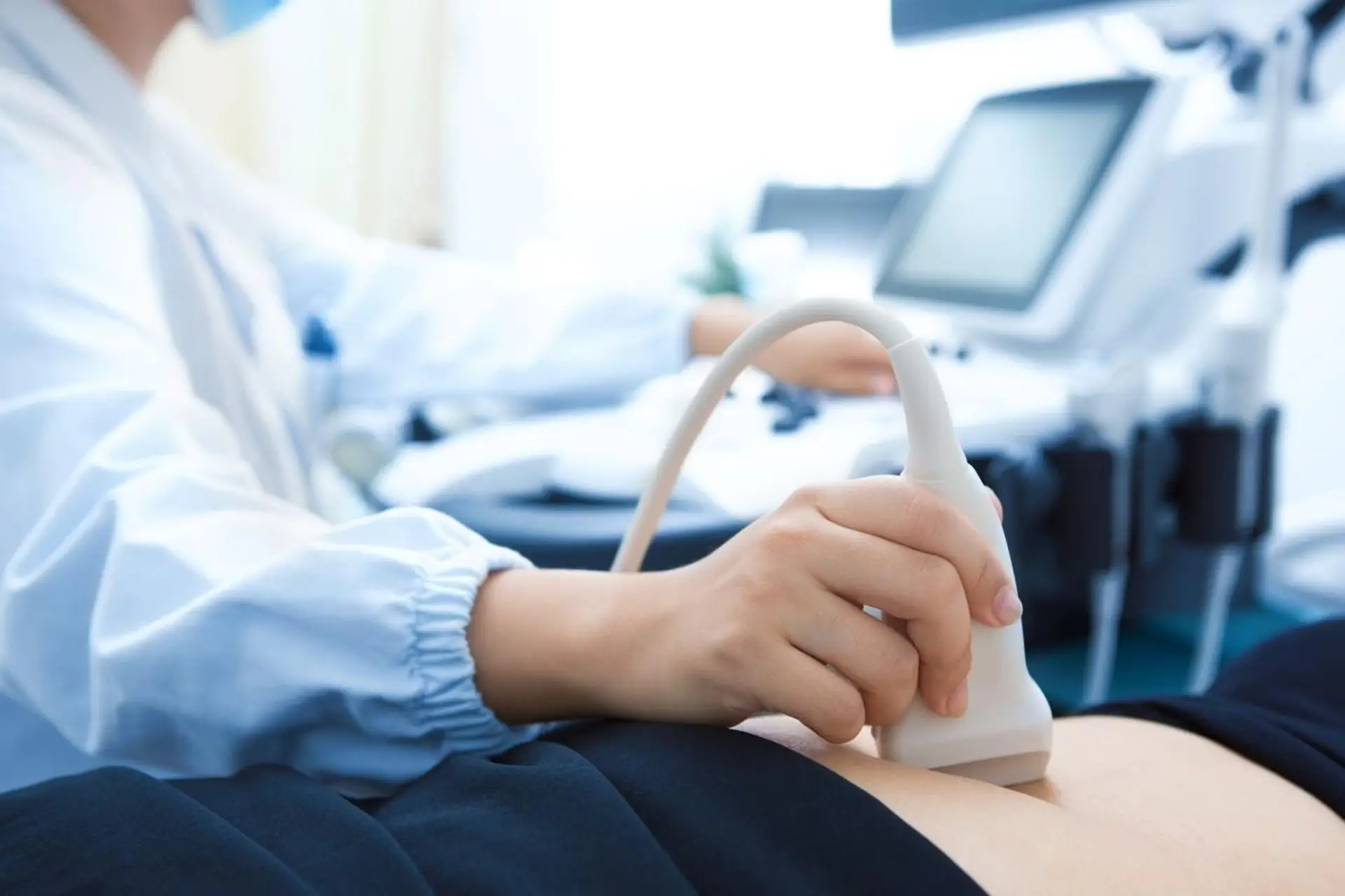
Diagnostic ultrasound uses returning sound echoes to create images. The sonographer changes the probe angle and position to examine a structure from several directions and may ask you to turn or briefly change your breathing. Gentle pressure and gel help obtain contact and useful views. The examination is therefore an active collection of information, not a single still picture. Some images are stored with measurements while other observations are made in real time. The amount of probe movement or the number of saved pictures does not reliably indicate whether the examination is normal or abnormal.
Ordinary gray-scale images describe the appearance of structures. Doppler techniques add information about movement of blood and can display direction or measure aspects of flow. Colors on the screen represent settings used for that assessment; red and blue should not be read as a simple healthy-versus-unhealthy code. A vascular examination has a specific question and protocol. It is different from assuming that every ultrasound includes a complete vessel assessment. If the clinical concern involves a clot, narrowing or another circulation issue, the request needs to identify the relevant vascular examination and body region.
An abdominal ultrasound and a pelvic ultrasound can have different preparation because the sound needs a useful route to the structures being examined. Bowel gas can obstruct that route. For some pelvic examinations, a filled bladder can improve the view from the abdomen, while another approach may use different instructions. If an internal examination is considered, the team should explain the approach and discuss consent and comfort. General information about an approach is not confirmation that every type is offered locally. Follow the instructions for the agreed examination, and clarify any conflict before changing food, fluid or medication routines.
Sound does not travel through every part of the body equally well. Air, bone, depth and the available viewing position can limit what is seen. A report that says a structure was not well visualized is different from a report saying that it was examined and appeared normal. Ask whether the examination answered the main question or whether another method is needed. Depending on that question, the next step could involve clinical follow-up, additional ultrasound views, MRI or CT. A recommendation for another test can reflect a limitation of the view rather than a serious diagnosis.
| Focus | What the study can add | What to keep in mind |
|---|---|---|
| Abdominal organs | Sound-wave imaging can assess selected abdominal organs. | Bowel gas and other factors can limit the structures seen. |
| Pelvic structures | The ordered approach determines how pelvic organs are examined. | Bladder instructions and consent discussions depend on the approach. |
| Blood flow | A Doppler examination can assess aspects of blood flow. | Vascular studies require their own protocol and availability confirmation. |
A doctor’s order is required before your scan. Preparation varies by examination. Some studies require fasting; others require a full bladder. Ask for instructions for the exact body area on your order and confirm how to take regular medicines.
An order for an abdominal, pelvic or vascular examination needs more detail than “ultrasound.” Tell scheduling the exact wording, the body region and any additional instructions. Confirm that the requested examination is available at your preferred center. An educational description of a type of ultrasound is not confirmation that every protocol is offered at every location.
Ask whether you need to avoid food, drink water or arrive with a full bladder for your study. Obtain the amount and timing directly from the imaging team. If preparation is difficult because of medication or another health condition, call for individualized instructions instead of changing the plan yourself.
For an estimate, provide the complete ultrasound order so the office can identify the study being quoted. Ask whether interpretation is included and who will send the report to your clinician. Before leaving, confirm how to arrange your results discussion and request copies if another office needs them.
Ask the team to explain the approach for the ordered examination. Follow the positioning and preparation instructions, and raise questions about comfort or consent before the examination proceeds.
Your radiologist’s report goes to the ordering clinician, who will explain the findings and next steps. Contact our records team for copies of your images or report.
Request images and reportsRequest an estimate for the examination on your referral, including contrast and radiologist interpretation. Confirm your plan’s network status and authorization requirements.
Costs, insurance & financingAvailable locations: Richmond Avenue, Walters Road, Dickinson. Confirm your exact protocol when booking.
View locations & hoursCommon questions
Read the complete examination name and body region from your order to scheduling. Our team will confirm availability, the location and the instructions for that diagnostic study.
Call 877-345-4244 with your order or request a callback. The team confirms the requested protocol, appropriate center and appointment options. A callback request does not itself confirm a booking.
Ask for a current quote for the exact study, including any contrast and the radiologist’s interpretation. Confirm separate charges and payment terms. If using insurance, check your plan’s network, benefits and authorization requirements.
Read each requested study to scheduling. Ask whether separate appointments or estimates are needed and whether any preparation instructions conflict. The office can obtain clarification from the referring clinician when necessary.
Ask which earlier images and reports are useful and how to transfer them. An existing scan may help comparison, but the clinical team determines whether it answers the current question or meets any specialized analysis requirements.
Arrange the report discussion with the clinician who ordered the examination or manages your care. Ask how the report will reach that office and when follow-up is appropriate. Records staff can help arrange copies for another provider.
Still images document measurements and appearances, while moving images can record changing structures or flow during the examination. These are complementary ways of recording findings. The examination method depends on the clinical question; the presence of video clips does not itself identify a problem.
No. Coverage is defined by the examination performed. Abdominal and pelvic studies have different targets and may require different approaches. Check the examination name and report before assuming an organ was included, especially when discussing symptoms in a different body region.
Yes. Advantis requires a doctor’s order for every scan, including self-pay appointments. You can request a callback before you have the order; it must be available before the examination.
Have your referral ready. Our team will confirm your scan, location and available appointment times.
The patient guide
Preparing for a scan
Fasting and bladder instructions depend on the examination. Confirm the exact ultrasound on your order before preparing.
Costs & appointments
Looking for affordable ultrasound options without insurance? Compare the complete self-pay estimate and ask Advantis about its Best Price Guarantee before booking.
Preparing for a scan
Prepare for an abdominal ultrasound with clear questions about fasting, water, medicines and the exact examination on your doctor’s order.
Preparing for a scan
Understand pelvic ultrasound preparation, full-bladder instructions and questions to ask about abdominal and internal examination approaches.
Understanding imaging
Understand how ultrasound images become a report, who explains the findings and what to do if you have not received your results.
X-ray & ultrasound
Doppler ultrasound adds information about blood flow. Learn what vascular orders mean and how preparation depends on the area examined.
