
The short answer
Conventional ultrasound shows tissue structures using sound waves. Doppler techniques add information about blood movement, and a duplex examination combines structural and flow assessment. The body area and clinical question determine the requested protocol and preparation.
- Doppler is a technique used within an ultrasound examination.
- Arterial and venous studies answer different questions.
- The order should identify the region and side when relevant.
- Preparation depends on the specific vascular study.
- Sudden severe symptoms need medical assessment, not routine scheduling.
| Term | Meaning to clarify |
|---|---|
| Conventional ultrasound | Structural imaging with sound waves |
| Doppler | Assessment of blood movement |
| Duplex | Structural and flow assessment combined |
| Arterial or venous | Which type of vessel the order targets |
| Unilateral or bilateral | One side or both sides |
What Doppler adds to the images
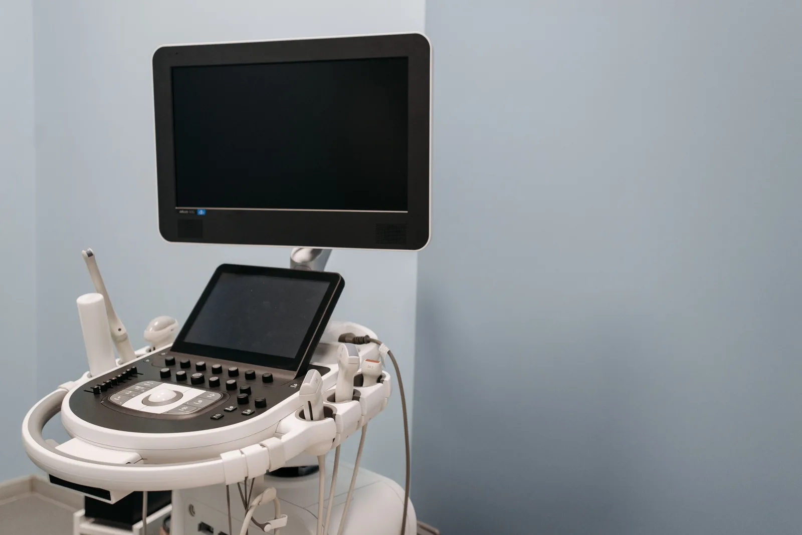
Ultrasound can create real-time images of structures. Doppler techniques use information from reflected sound to assess blood movement; flow may be displayed using color or a waveform. A duplex study combines structural imaging with Doppler assessment. The technologist acquires the requested views and measurements, and a qualified clinician interprets them. The colors are display conventions, not a simple red-is-bad or blue-is-good result for a patient to interpret independently.
Why the exact vascular order matters
A venous study of a leg is different from an arterial assessment of that leg or a carotid study in the neck. Some orders are unilateral and others bilateral. Read the full order to scheduling and ask whether it requires clarification. Avoid booking a general soft-tissue ultrasound when the clinician requested a vascular protocol. The right terminology helps the center reserve the correct examination and provide an accurate estimate.
Grayscale, color and spectral images have different jobs
Conventional grayscale ultrasound displays tissue structure. Color Doppler adds information about detected blood flow, and spectral Doppler represents flow measurements as a waveform over time. These displays can be used together during one vascular examination. They are complementary ways of assessing a clinical question rather than separate quality levels.
The color on the screen is not a simple red-for-healthy, blue-for-unhealthy code. Its meaning depends on the equipment settings and flow direction being displayed. Ask the clinician to explain the interpreted report rather than drawing conclusions from a brief view of the monitor. The sonographer may adjust the display repeatedly while obtaining measurements.
Arterial and venous ultrasound are not interchangeable
Arteries carry blood away from the heart and veins return it. An arterial study and a venous study may therefore evaluate different problems even when both involve the same leg. The order needs to identify the vessels, side and clinical question. A carotid examination, an abdominal vascular study and a leg vein examination also have different requirements.
When contacting Advantis, read the specific examination and ask the team to confirm availability for that vascular protocol and center. General ultrasound availability does not establish that every vascular study is performed at every location. If the order changes after booking, update the center so the appointment length and preparation can be checked.
Prepare for contact, positioning and a results conversation
The examination uses a probe and gel on the skin over the area being assessed. The sonographer may need to reposition you or apply pressure as part of the ordered study. Tell staff about tenderness, limited movement or dressings before the examination begins so the approach can be discussed.
Ask your referring clinician when the result should be reviewed and whether the clinical concern requires urgent assessment. Do not wait for a routine ultrasound appointment if you develop severe or rapidly worsening symptoms. For a planned study, keep the full order and earlier relevant reports together and confirm who will receive the final interpretation.
Ask for preparation specific to the body area
An examination of neck or limb vessels can have different preparation from an abdominal vascular study. Ask whether fasting, clothing choices or arrival arrangements matter for your appointment. If you have several tests booked together, confirm which instructions apply to each one. Do not assume that a full-bladder instruction from an earlier pelvic scan applies to a vascular examination. Contact scheduling when instructions are missing or contradictory.
During the examination
The technologist applies gel and moves a probe over the area being examined. Positioning and gentle pressure may be needed to obtain the requested information. Tell the team if the area is painful, movement is limited or you need assistance changing position. You may hear sounds generated from flow measurements. Ask about the examination process, but avoid interpreting time spent on one area or the technologist's expression as a diagnosis.
Confirm the requested study with Advantis
Advantis lists diagnostic ultrasound at its Richmond Avenue, Walters Road and Dickinson centers. Call to confirm whether the specific arterial or venous protocol on your order is available and at which location. Give the side and region, and ask whether prior studies should be transferred for comparison. A doctor's order is required. A general ultrasound listing should not be taken as confirmation of every specialized vascular study at every center.
Arrange a results discussion
Ask which clinician will receive and explain the report and whether any follow-up is expected. If your symptoms are sudden or severe, contact a medical professional promptly rather than choosing a routine scan yourself. An outpatient callback form does not assess urgency. For a planned follow-up examination, keep previous reports and the clinician's recommended interval so the new study can be compared in the correct clinical context.
Your Next-Step Checklist
- Confirm arterial versus venous, body region and side.
- Ask whether the exact vascular protocol is available.
- Follow preparation instructions for that study only.
- Use the report, not the screen colors, for the results discussion.
Preparing for different ultrasound examinations
Getting your ultrasound results
Frequently Asked Questions
Does Doppler ultrasound use radiation?
Diagnostic ultrasound uses sound waves rather than ionizing radiation. Follow the preparation instructions for your particular study.
Can I request any Doppler scan at any center?
Confirm the specific protocol and location with scheduling. The word Doppler alone does not identify a complete examination.
Will the technologist tell me the diagnosis?
Ask who provides the interpreted report and how your ordering clinician will discuss it. Image acquisition and the clinical results conversation are different steps.
Does blue on Doppler mean there is no oxygen in the blood?
No. The displayed colors depend on Doppler settings and flow direction. They should not be interpreted as a simple oxygen-level display.
Can a general ultrasound appointment cover any Doppler test?
Not automatically. The center must confirm the exact vascular protocol, equipment and appointment arrangements on your order.
General educational information. Your examination and preparation depend on your clinical needs. Follow the instructions provided for your appointment.
Further reading: RadiologyInfo: Vascular ultrasound