The staff is Amazing especially the MRI Tech she was patient and helpful due to my anxiety.
Ciairra Washington
 Google review
Specialized imaging
Detailed Small Bowel Imaging | Board-Certified Radiologists
Best Price Guarantee 4.9
4.9Patient stories
Real patient stories from Advantis MRI & Diagnostic Imaging.
4.9 / 5from 1,000+ reviewsThe staff is Amazing especially the MRI Tech she was patient and helpful due to my anxiety.
Google reviewGreat customer service and gave me really good comfort while getting my MRI. I will recommend coming here for services.
Google reviewThey work to make sure things are comfortable and correct. Great place to get an MRI, if you have to.
Google reviewVery clean and neat lobby and they have snacks!! Staff was wonderful and the tech was really quick and generous.
Google reviewSpeedy service, the staff was very professional and knowledgeable.
Google reviewThe entire visit was amazing. The staff were kind and personable, and I really appreciated how helpful and accommodating everyone was.
Google reviewUnderstand the examination
Enterography provides detailed images of the small intestine. CT enterography commonly involves drinking a liquid and receiving intravenous contrast. MR enterography is a different technique, so the name and modality on the order matter.
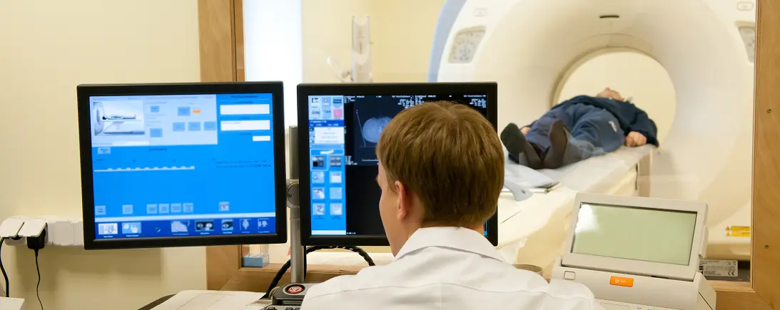
Enterography is tailored to the small bowel rather than being a generic abdominal examination. In CT enterography, liquid taken by mouth helps distend the bowel and intravenous contrast helps assess tissues during the CT acquisition. The images can show the bowel wall and surrounding structures, which can be useful when investigating inflammation, narrowing or complications such as an abscess or fistula. This approach provides information that is different from simply looking at the inner surface of the bowel. The examination name on the referral matters because a standard abdominal CT may use a different preparation and imaging plan for a different clinical question.
MR enterography uses MRI rather than CT to examine the small bowel. It can also involve oral preparation and intravenous contrast, but the image acquisition, safety screening and experience in the scanner differ. MRI does not use ionizing radiation, which may be relevant when a clinician expects repeated bowel imaging. That does not make it the right choice for every circumstance. The clinical question, ability to complete the examination and MRI suitability all influence the decision. “Enterography” alone does not establish which method has been requested. Clarify CT versus MR before comparing preparation advice or assuming that one appointment can substitute for the other.
A diagnosis such as Crohn’s disease is based on more than a single scan. Clinical history, examination, blood or stool tests, endoscopy and imaging can contribute different pieces of information. Endoscopy allows a clinician to inspect the lining and obtain tissue samples, while enterography helps assess bowel structures and areas around them. These methods can be complementary. When results are discussed, ask which part of the diagnostic question the scan answered and whether any tissue or laboratory information is still needed. A useful follow-up explains the findings in plain language and connects them with symptoms, rather than treating an imaging label as the entire diagnosis.
In Crohn’s disease, imaging can help assess the distribution of bowel involvement and look for complications. A report may discuss inflammation, a narrowed segment, an abnormal connection between structures or a collection outside the bowel. These observations are not interchangeable, and their clinical significance depends on the surrounding history and other tests. Explain previous bowel operations, established disease and the way symptoms have changed, so the interpreting team can relate the images to the right question. When reviewing the report, ask whether the main finding concerns the bowel wall, passage through a narrowed segment or a complication outside it. That distinction helps make a discussion about further investigation or treatment more understandable.
A small-bowel follow-through uses X-ray imaging to observe contrast moving through the small intestine. CT and MR enterography use different methods to assess the bowel and surrounding structures. The tests may sound similar because they all concern the small bowel, but their preparation, images and diagnostic strengths differ. In particular, the choice affects whether the clinician is examining passage through the bowel, cross-sectional anatomy or complications around the bowel wall. If an older report mentions a follow-through, bring it to the clinical discussion without assuming that a newly requested enterography will repeat the same examination. Ask what information is already available and what the new method is expected to add.
MR enterography requires a patient to remain sufficiently still during several image acquisitions, sometimes following breathing instructions. This is part of obtaining images that can be interpreted reliably. Explain difficulty lying comfortably, anxiety or an inability to follow a requested breath-hold. The team can discuss the practical requirements of the examination. A referral for MR enterography also requires MRI safety screening; the oral preparation does not remove the need to review implanted devices and other MRI-related considerations.
| Focus | What the study can add | What to keep in mind |
|---|---|---|
| Small bowel | Enterography uses a specialized approach to examine the small intestine. | CT and MR enterography are different examinations. |
| Bowel preparation | The imaging protocol can include oral preparation and contrast steps. | Obtain instructions for the confirmed modality and appointment. |
| Clinical purpose | The order may ask about inflammation or another defined bowel concern. | The report is considered with the wider gastrointestinal assessment. |
A doctor’s order is required before your scan. Confirm whether your clinician ordered CT or MR enterography and which is available. Ask when to arrive for oral preparation, what you may eat or drink, and whether kidney function results are needed.
The team confirms the examination and explains the protocol-specific steps. For specialized imaging or analysis, ask what will be performed, which records are needed and who will interpret the result.
Your radiologist’s report goes to the ordering clinician, who will explain the findings and next steps. Contact our records team for copies of your images or report.
Request images and reportsRequest an estimate for the examination on your referral, including contrast and radiologist interpretation. Confirm your plan’s network status and authorization requirements.
Costs, insurance & financingCall with your referral and preferred location to confirm where your examination is available.
View locations & hoursCommon questions
No. Enterography uses a specific preparation and imaging approach for the bowel. Tell scheduling the complete examination name.
Call 877-345-4244 with your order or request a callback. The team confirms the requested protocol, appropriate center and appointment options. A callback request does not itself confirm a booking.
Ask for a current quote for the exact study, including any contrast and the radiologist’s interpretation. Confirm separate charges and payment terms. If using insurance, check your plan’s network, benefits and authorization requirements.
Read each requested study to scheduling. Ask whether separate appointments or estimates are needed and whether any preparation instructions conflict. The office can obtain clarification from the referring clinician when necessary.
Ask which earlier images and reports are useful and how to transfer them. An existing scan may help comparison, but the clinical team determines whether it answers the current question or meets any specialized analysis requirements.
Arrange the report discussion with the clinician who ordered the examination or manages your care. Ask how the report will reach that office and when follow-up is appropriate. Records staff can help arrange copies for another provider.
Not automatically. Imaging can assess bowel structures and surrounding tissues, while endoscopy allows direct inspection and tissue sampling. Your clinician may need both kinds of information. Ask whether the purpose is to locate disease, assess complications, evaluate response or establish a diagnosis. That purpose helps explain why another investigation might remain necessary even after the enterography images have been reviewed.
The liquid preparation helps the bowel be assessed during the examination. If you cannot tolerate the planned drink or have difficulty completing it, tell the team rather than substituting another product or skipping the preparation. The instructions depend on the imaging method and clinical plan. An ordinary abdominal CT preparation should not be assumed to apply to a dedicated enterography examination.
No. The significance depends on the appearance, location, symptoms and established diagnosis. Imaging findings need to be considered with other clinical information. Ask the treating clinician whether the report changes the assessment of disease activity or identifies a complication needing a separate plan. Do not infer a treatment decision from the word “narrowing” alone.
No. The imaging method and the information sought also differ. A follow-through examines contrast passage with X-rays, while CT or MR enterography produces a different assessment of bowel structures. Similar preparation terminology can obscure those distinctions. Use the complete examination name when reviewing instructions or discussing the reason a particular study was selected.
Yes. Advantis requires a doctor’s order for every scan, including self-pay appointments. You can request a callback before you have the order; it must be available before the examination.
Have your referral ready. Our team will confirm your scan, location and available appointment times.
The patient guide
Vascular & specialized imaging
CT enterography uses a dedicated small-bowel protocol. Learn how it differs from routine abdominal CT and what to confirm before your visit.
Preparing for a scan
Use the instructions for your ordered CT study to plan food, medication, contrast and arrival time.
