The short answer
Diffusion tensor imaging is an MRI technique that examines patterns of water movement in tissue, often used to study white-matter organization. It can supply additional information, but it is not a stand-alone test that proves every brain injury or explains all symptoms.
- DTI requires a specific MRI acquisition and analysis.
- Tractography images are derived representations, not direct photographs of nerve fibers.
- Measurements need expert interpretation and quality review.
- A result alone does not establish the cause or timing of an injury.
- Confirm the requested protocol with the ordering clinician.
| Component | What it contributes |
|---|---|
| Conventional sequences | Anatomical and tissue information |
| Diffusion acquisition | Measurements of water-motion patterns |
| Tractography | A model derived from diffusion data |
| Clinical history | Context needed to interpret the findings |
| Specialist review | Assessment of relevance and limitations |
What is different from conventional MRI
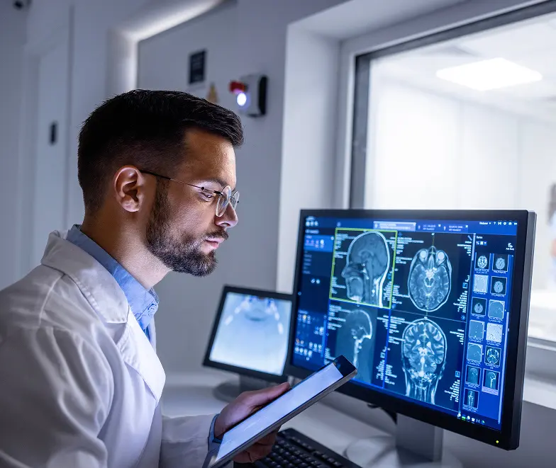
A conventional brain MRI primarily provides images of anatomy using several sequences. DTI adds measurements related to the direction of water diffusion. These can help characterize white-matter organization and support specialized assessment. The colorful fiber maps sometimes associated with DTI are computer-derived representations. They should not be read as a direct photograph of every connection or as a simple normal-versus-abnormal answer to a patient's symptoms.
What the result cannot establish on its own
DTI findings need to be considered with the injury history, clinical examination and other information. Technical factors and biological variation can affect measurements. An unusual value does not independently prove the mechanism, date or legal cause of an injury. Similarly, an unremarkable study does not mean symptoms are imaginary. Ask the clinician what question the examination is intended to answer and how a possible result would change the assessment or care plan.
Understanding diffusion maps and tractography
Diffusion imaging evaluates the movement of water within tissue. Diffusion tensor techniques characterize directional patterns and can produce maps used to study white-matter pathways. Tractography is a computational representation based on the acquired data; its colored pathways should not be mistaken for a direct photograph of individual nerve fibers.
This distinction is useful when a striking image appears more definitive than the accompanying explanation. Ask what was measured, which question the analysis was intended to address and whether the interpreting clinician identified limitations. A visually unusual map is not enough on its own to determine the cause or timing of an injury.
Keep research findings separate from an individual diagnosis
A technique can show differences between research groups without providing a standalone answer for every individual patient. Interpretation may depend on acquisition, analysis choices and the clinical setting. If DTI is being requested after an injury, the clinician still needs the history, examination, conventional imaging and other relevant assessment.
Ask how the proposed examination will influence your care. If a report is needed for a specific specialist, confirm that it will contain the measurements and interpretation that specialist expects. Do not rely on a general promise that a scan can prove every symptom or precisely date an event. The limits of the technique should be explained before the examination is purchased.
Organize a DTI referral before choosing the appointment
Advantis lists DTI arrangements at Richmond Avenue and Dickinson, rather than Walters Road open MRI. Read the exact request to scheduling and identify the referring clinician. Ask whether a routine brain MRI is included, which analysis is requested and how the results will be delivered.
Collect previous brain imaging and relevant reports so the team can advise on comparison requirements. If implants, anxiety or difficulty remaining still could affect the visit, discuss them before selecting a center. Keep a copy of the agreed examination details with your estimate; an ordinary brain MRI appointment should not be assumed to include specialized diffusion analysis.
Make the referral specific
Before scheduling, ask whether the order requests routine brain MRI with DTI, a separate analysis or another protocol. If a specialist needs particular sequences or report outputs, share those requirements with the imaging center. A general brain MRI booking may not include the requested acquisition. Clarifying this beforehand is more useful than discovering after the appointment that the referring office needs data that were never collected.
Location and safety planning
Current Advantis scheduling guidance places DTI requests with the high-field MRI locations at Richmond Avenue or Dickinson, subject to protocol confirmation. It should not be assumed available on the Walters Road open scanner. Tell the team about implants, retained metal and difficulty remaining still. Ask for any contrast instructions relevant to the actual order. A doctor's order is required, and completing online screening questions does not replace the team's MRI safety assessment.
Ask what is included in the estimate
Clarify whether the quoted amount includes the MRI acquisition, radiologist interpretation, DTI processing and any separate report expected by your clinician. If another organization will analyze the images, ask who arranges the transfer and whether its fee is separate. Keep these arrangements documented with the referral. An advanced-sounding service name is not enough to determine what files and reports will actually be delivered after the appointment.
Keep the full record for clinical review
Request instructions for obtaining the images and all associated reports. If a different specialist will review them, ask which formats are needed and where to send them securely. At follow-up, ask how the findings relate to the history and which uncertainties remain. Avoid making treatment or legal conclusions from a single colored image. The appropriate interpretation comes from a clinician reviewing the complete evidence rather than the appearance of one visualization.
Your Next-Step Checklist
- Identify the clinical question and requested analysis.
- Ask how DTI will be interpreted with conventional imaging.
- Confirm Richmond Avenue or Dickinson arrangements.
- Keep the complete images, radiology report and analysis together.
How quantitative brain reports differ
Frequently Asked Questions
Does DTI prove a concussion?
It is not a stand-alone proof of concussion or its cause. Diagnosis and interpretation require the clinical history and appropriate assessment.
Can I request DTI at any MRI location?
Confirm availability for the actual protocol. Advantis guidance does not place DTI on the Walters Road open scanner.
Is DTI the same as NeuroQuant?
No. DTI evaluates diffusion-related information, while NeuroQuant provides volume measurements from suitable MRI data. They answer different questions.
Are the colored pathways a photograph of individual nerves?
No. Tractography is a computed representation based on diffusion data. The result needs interpretation within the technical and clinical limits of the method.
Does every brain MRI include DTI analysis?
No. Specialized acquisition and analysis need to be requested and confirmed. Ask scheduling to review the exact order before booking.
General educational information. Your examination and preparation depend on your clinical needs. Follow the instructions provided for your appointment.
Further reading: NIH: how MRI works · NINDS: Traumatic brain injury research