The short answer
Brain MRI can answer a specific neurological imaging question. The order, contrast protocol, prior studies and follow-up plan shape the visit.
- A focused pituitary or vascular question may need a different protocol.
- Have earlier brain studies available when requested.
- Discuss the report with the clinician responsible for your care.
| On your order | What to clarify |
|---|---|
| Brain MRI | Brain tissue and the ordered focused assessment. |
| MRA | Blood-vessel imaging; a separate request. |
| Head CT | A different imaging method and protocol. |
Routine brain imaging and vascular imaging answer different questions
Brain MRI can investigate brain tissue and related structures in the assessment of tumors, inflammation, stroke-related injury, seizure causes and other neurological concerns. A related examination, MR angiography, focuses on blood vessels. These studies can complement one another, but the names are not interchangeable. A focused question about the pituitary gland or another small structure may also need tailored images. This is why the clinical history and complete order matter. A general brain MRI is not automatically a comprehensive test of every neurological function or every vessel in the head.
Read the report as a structured explanation
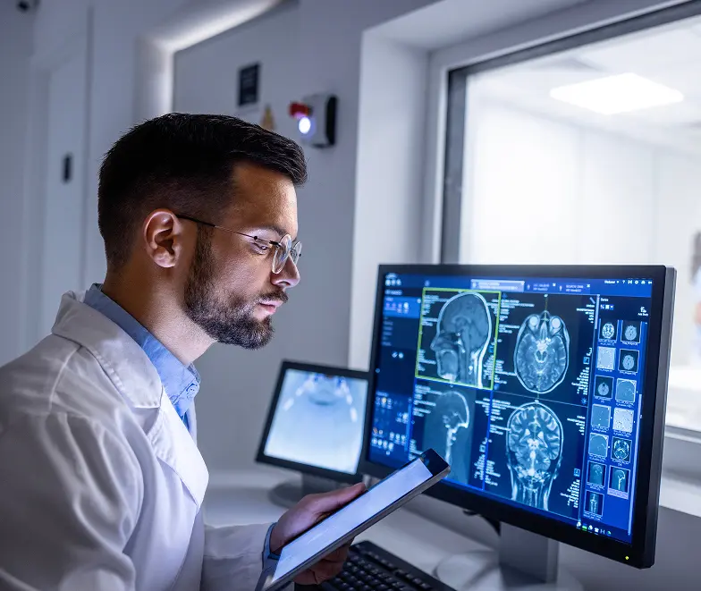
A brain MRI report commonly records the reason for the examination, the technique, previous studies used for comparison, the findings and an impression. The impression highlights the conclusions the radiologist wants the referring clinician to consider. The comparison section can be especially useful: a finding that is unchanged from earlier imaging may be interpreted differently from something new. Some observations are incidental and do not explain the symptom that prompted the scan. Ask which findings matter for your care and whether any recommendation depends on obtaining older images from another facility.
Why the examination includes several image sequences
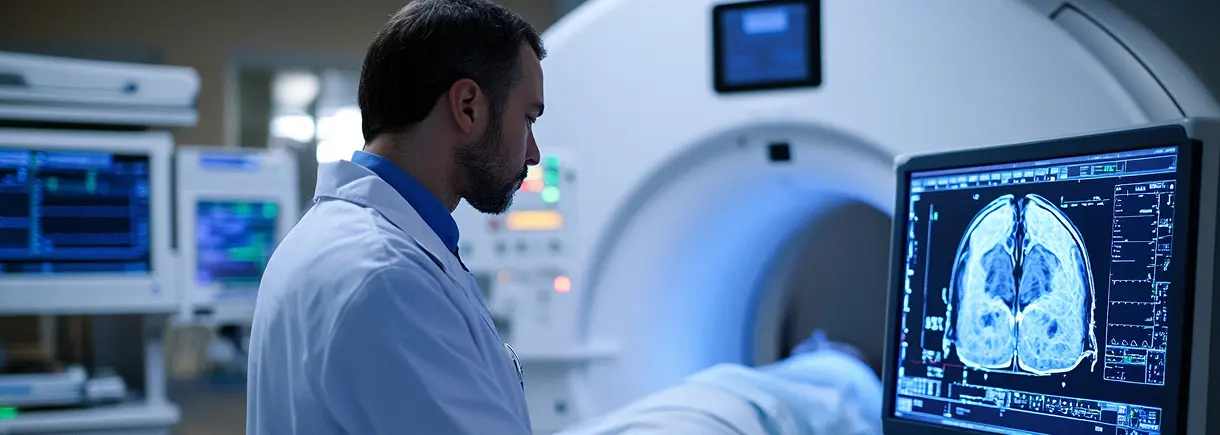
A brain MRI uses different sequences to show different tissue characteristics. You may notice changes in scanner sounds or short pauses between them. The examination is not simply one long photograph, and staying still through each sequence helps the team obtain useful images.
Ask the technologist how to communicate if you are uncomfortable. Positioning equipment around the head helps acquire the images and limits movement, but it can feel unfamiliar. Mention anxiety, pain or difficulty lying flat before the appointment so the team can discuss the practical requirements of your ordered study.
Plan how to understand the report
The radiologist's observations and impression answer the imaging question, while your clinician connects those findings with symptoms, examination and other tests. A report may mention findings that are not the main explanation for your concern. Ask which observations are relevant and whether comparison with earlier imaging changes the interpretation.
If the report recommends another examination, ask what additional question it would answer and how soon it is needed. Arrange the results conversation before assuming that a normal scan ends the assessment or that an unfamiliar phrase establishes a diagnosis.
When the question is specifically about blood vessels
MR angiography can examine vessels in a chosen region using MRI techniques. Depending on the question and method, it may be performed with or without injected contrast. It can help assess abnormalities such as narrowing or an aneurysm, but the vascular territory must be specified. An MRA of the head and an MRA of the neck are different requests, and a routine brain MRI should not be assumed to include either. If the referral mentions more than one examination, ask the ordering office what each is intended to evaluate. This distinction explains why the complete order matters even when all the studies are commonly described as an MRI of the head.
What does a brain MRI show?
A brain MRI produces detailed images of brain tissue without ionizing radiation. Clinicians may use it to investigate conditions such as tumors, inflammation, seizure-related changes or damage from a stroke. The requested technique depends on the clinical question. A brain MRI and an MRA of the blood vessels are different studies; one should not be substituted for the other without clarification.
How long does a brain MRI take?
There is no single duration for every head MRI. The imaging sequences, whether contrast is needed and your ability to remain still affect the examination. Ask scheduling for the expected scanner time for your order and the full arrival-to-departure window. If you also have another body area scheduled, check whether the estimate includes both studies.
What a brain MRI can investigate
Brain MRI can help evaluate structural abnormalities, some neurological conditions and changes already seen on earlier imaging. Examples include assessment of tumors, inflammation, stroke-related changes or a focused pituitary concern. These are uses of the technique, not a reason for everyone to have screening. The clinical history and examination determine whether MRI is appropriate and which protocol is needed. A focused question may require additional sequences or a separate vascular study.
Reading the report without jumping to a diagnosis
The report describes the images and the radiologist’s interpretation of them. A finding may be relevant to the concern being investigated, incidental or something that needs comparison with an earlier study. Ask the ordering clinician which findings matter and what happens next. If the report recommends another examination, ask what question that would answer. The need for follow-up does not, by itself, establish a particular diagnosis.
Will contrast be needed?
Some brain examinations use an injected contrast agent; others do not. Your clinical team determines the protocol. Ask scheduling to check the wording of your order and tell them about previous contrast reactions, kidney conditions or possible pregnancy. Avoid using another patient’s experience to decide which type of scan to request.
Prepare the practical details
Keep your order, referring office’s number and any implant identification information together. Ask whether previous brain imaging should be supplied and how to send it. Discuss concerns about positioning or enclosed spaces before booking. Write down your arrival time, center address and any food, clothing or medication instructions for this appointment.
Plan how you will receive results
Ask the ordering office when and how the report will be discussed. Write down the question your clinician wants imaging to answer so you can return to it during follow-up. If another specialist needs the images, ask about the release process. A routine outpatient appointment is not an emergency evaluation; call 911 for a suspected medical emergency.
Ask Advantis about your order
Call with the full requested study to confirm protocol availability and the appropriate center. Advantis lists high-field MRI at Richmond Avenue and Dickinson, and open MRI at Walters Road. The scheduling team needs to review your particular examination before confirming a location. Ask for an estimate covering the ordered study and any contrast.
Your Next-Step Checklist
- Confirm the exact brain protocol and contrast plan.
- Raise positioning and implant questions early.
- Ask how to communicate during the scan.
- Arrange a clinician-led results discussion.
Brain MRI in Houston and Dickinson
Frequently Asked Questions
Is brain MRI the same as an MRA?
Brain MRI examines the requested brain structures. MRA focuses on blood vessels and may require a separate order. Read both study names to scheduling if both are requested.
Why might earlier brain images be needed?
A comparison may help determine whether a finding has changed. Ask how to transfer both images and reports from another provider before the examination.
Can movement or metal affect brain MRI images?
Yes. Movement can blur images, and some metal can distort them. Tell the team about implants or earlier surgery and explain if pain, tremor or anxiety makes remaining still difficult. These issues can affect planning and image quality as well as the safety review before scanning.
Does every unfamiliar phrase in a brain MRI report mean disease?
No. Reports use technical language and may describe common or incidental observations. Read the impression with your clinician and ask how it relates to the reason for imaging. Prior studies, age, symptoms and other findings can change the significance of a description that sounds concerning on its own.
Does MRA always require contrast?
No. Some MRA techniques can show blood vessels without an injection, while others use contrast. The choice depends on the region and clinical question. Ask which examination has been requested and how it relates to any routine brain MRI included in the plan.
General educational information. Your examination and preparation depend on your clinical needs. Follow the instructions provided for your appointment.
Further reading: RadiologyInfo: brain MRI · RadiologyInfo: article brain mri report · RadiologyInfo: MR angiography